Phan Thi Hien, "Esophagus-stomach-duodenum endoscopy in children", Medical Publishing House, Hanoi, Jan 2019 (page 181-186). Extract from specialty book: “NỘI SOI THỰC QUẢN – DẠ DÀY – TÁ TRÀNG TRẺ EM”, Nhà xuất bản y học, Hà Nội, ISBN: 978-604-66-3546-8.
INTRODUCTION
At the beginning of 1980, Ponsky and Gauderer had developed percutaneous endoscopic gastrostomy, opening a new approach in pediatric related to enteral feeding, especially in children with dysphagia, or inability of swallowing. This is a minimal invasive intervention that helps to optimize nutrition and easy care [1]. The advantages of percutaneous endoscopic gastrostomycomparing to surgery include short technique, low price, smallerincision, short length of lesion, decreasing reflux and complications after surgery (inflammation, intestinal adhesion, intestinal obstruction, abdominal pain, intestinal movement disorders) [2].
1. INDICATIONS
Percutaneous endoscopic gastrostomyis indicated if without history of abdominal surgery. The aim of gastrostomy is for delivering drugs, foods or reducing stomach pressure. This technique does not affect Nissen fundoplication, pyloroplasty surgery or pyloromyotomy in the future (if needed). Specific indications:
- Gastrostomy indication for feeding in cases with severe reflux or neurological defect, myopathies, cystic fibrosis and chronic respiratory failure. Sometime gastrostomy combines withNissen fundoplicationfor avoiding reflux in each specific case [1].
- Some special gastrostomy indications in cases who need jejunal feeding (Cosanum, Schilieren, Switzeland). This technique is indicated when patients have severe reflux, weak ability to make stomach empty or chronic pancreatitis [1].
2. CONTRAINDICATIONS
2.1. Absolute contraindications
- Unsuccessful transillumination of gastric wall on endoscopy.
- Suspect the anterior wall of the stomach is not opposed to the abdominal wall because transverse colon or other organs in the abdomen.
- The anterior wall of the stomach is not opposed to the abdominal wall due to liquid of abdomen or other reasons [2].
- Coagulopathy.
2.2. Relative contraindications
- Ascites, peritoneal dialysis, active gastritis and or peptic ulcer [1,2].
- Portal hypertension includes stomach varices [1] and abdominal tumor [2].
- Abnormal anatomy includes intestinal malrotation or colon, spleen, liver interfere between stomach and abdominal wall, hepatosplenomegaly, stomach in chest, gastrectomy and abdominal surgery [1,3].
-Hepatosplenomegaly, scoliosis, obesity, ventriculoperitoneal shunt (high risk of peritonitis due to fungus), patients younger 12 months or under 3kg of body weight [3].
These cases above are difficult to perform percutaneous endoscopic gastrostomy or cannot be to place or have high risk of complications because incorrect placement or severe inflammation,so should not perform this technique.
3. COMPLICATIONS
In children, severe complications were recorded around 3% cases with gastrostomy [1,3]. Always be careful with aspiration syndrome (because pumbingover amount of food by gastrostomy tube lead to reflux and choking), besides that bronchopneumopathy is the most common, other complications can happen. To limit complications of gastrostomy,abdominal ultrasound and intestinal transit X-rays are neededto assess the status of organs in the abdomen such as: hepatosplenomegaly, malrotation…[4].
3.1. Buried bumper syndrome
This complication is common, a consequence of excessive traction on the gastrostomy tube for a long time [1] or after the replacement of tube and may lead to gastric mucosal necrosis [3]. So, on endoscopy,the bumper is partially or not seen, buried in the gastric wall by mucosa [1, 3]. To prevent complication:
Avoiding excessive traction by carefully fixed gastrostomy tube, smooth feeding, without pulling the tube. If suspicion of traction on the gastrostomy tube, I need to perform endoscopy early.
3.2. Peritonitis
Peritonitis is a consequence of the separation between stomach and abdominal wall, tube malposition or leakage [1, 3]. Peritonitis can happen immediately after placement of tube or at time of replacing tube [1]. To prevent complication:
- Pumping maximum air in the stomach during insert trocar into the stomach as well as the process of passing the guide wire.
- Check carefully the bumper and external placement of the gastrostomy tube located inside stomach and outside abdominal wall after insertion or replacement [3, 4].
3.3. Gastrocolic fistula
Gastrocolic fistula occurs commonly in children with a history of abdominal surgery, and or weak transillumination of gastric wall [3]. To prevent complication:
- Position of patient head is slightly high to make the transverse colon go down [3].
- Check carefully for indication and contraindication, this is of crucial importance.
- Determine the position for trocar insertion based on steps.
This complication is usually relieved after withdrawing the gastrostomy tube several days [3].
3.4. Necrotising Fasciitis
This complication is rare, but severe due to skin infected at gastrotomy located, then necrosis and ulcer happens [1, 3].So, need to check carefully skin status and early infectious treatment. When necrotizing fasciitis appear, surgeon consultation is required.
3.5. Bleeding
Bleeding complication can happen due to vascular lesion on parietal stomal wall and can lead to death if it is not detected early [1]. Clinical symptoms are often discreet, therefore, needed to closely monitoring general condition of patient, anemia and, consultesurgeons when this complication is suspected.
3.6. Less severe complications
Complications are not severe account for around 7-13% of all cases:
- Common complication is an inflammation at gastrotomy located (this complication decreases due to antibiotic prophylaxis, standard skin disinfectants the mouth and throat area and the connected point between tube and wire before pulling from mouth into stomach, the length of cutting skin adequate is necessary because too narrow leads to anemia skin around tube, fixed ring outside only slightly touch abdominal wall, not tighten approach).
- Pneumoperitoneum is evidence of leaking liquid and air at around hole that is observed immediately in 1/3 cases because the abdominal wall and stomach are not close to each other (this complication reduces if the tube is slightly pulled). When having pneumoperitoneum, feeding must be delayed after 48-72 hours, and X-ray with contrast agent dissolving in water needs to be performed, if leaking into the peritoneal cavity, laparoscopy is needed [1, 3].
- Minor complications such as granulation reach up to 44%. When granuloma has developed immediately after insert gastrostomy tube, simple traction on the tube will abolish this development due to decreasing space between stomach and abdominal wall.The feeding needs to be delayed. Chemical treatment causes burning by topical application with silver nitrate (AgNO3).
- Local infection is usually serious because it can lead to septicaemia. Complication also can happen during the tube replacement process, the tube is not installed in the correct position, and endoscopy to detect this problem. Sometime, granuloma develops around the tube, also can lead to an opportunity of leaking bloody liquid [1].
4. TECHNIQUE
In pediatric, percutaneous endoscopic gastrostomyunder anesthesia usually performs quickly. A high risk of infection for diabetes patients. Common antibiotic prophylaxis are amoxicillin and cephalosporineintravenous injection [1]. Patients have to fast prior 8 hours, antibiotic prophylaxiscephalosporine (cefazolin 25mg/kg) or combination with amoxicillin-clavulanate 30 minutes prior gastrostomy and using twice at 8 hours and 16 hours. Blood testing, abdomen ultrasound and intestinal transit X-raybefore gastrostomy. Needed exact assessment swallowing process, excluded abnormal cardia and fundus position and other abnormal anatomy of gastrointestinal tract [3].
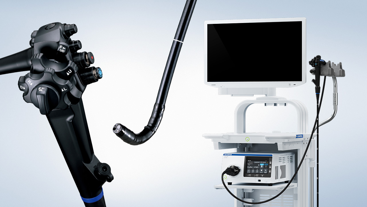
.jpg)
The percutaneous endoscopic gastrostomy kit
.jpg)
The gastrostomy tube for replacement
Common gastrostomy technique in pediatric is “pull through” technique. Implementing step:
- Patient position: supine,skin,mouth and throat disinfectant.
- Push endoscope tube into stomach and fully insufflate to approximate the stomach and the abdominal wall.The tip of endoscopeis controlled for application against the anterior wall of the stomach, to find exact for gastrostomy position base on transillumination of abdominal wall.The selected site is usually located on the anterior wall near junction of the body and the antrum (avoid cardia and ribs). The gastrostomy site needs to be perpendicular with the anterior wall of the stomach, should not locate in the lower part of the stomach to avoid risk of the colon or mesenteric entering stomach and abdominal wall. Using a finger push on the abdominal wall, where transillumination is clearly to determine the final stoma position [2,4].
- At the gastrostomy site, using a needle 25 gauge (1.5 inch) and syringe with lidocaine solution 1%to pass from skin into stomach until the air can be taken out, noticeably the needle always must be perpendicular with the abdominal wall. Lidocaine is injected with needle withdrawal if this needle is seen in the stomach.
.jpg)
Pump fully air and find the point of transillumination of abdominal wall
.jpg)
Determine the point to insert troca by finger imprints
.jpg)
Insert troca from abdominal wall into stomach
.jpg)
Use snareto pull the wire out of the mouth
.jpg)
Pull gastrostomy tube from mouth through abdominal wall
.jpg)
Internal Bumper in stomach
Use one troca following the direction of the needle into the stomach. At this time, the endoscopist needs to pump full air into the stomach to make the anterior gastric wall always pressed against the abdominal wall and to avoid interposition of spleen, liver, especially transverse colon.
- Guide wire passes through troca into stomach, snare passes through interventional channel of endoscope into stomach to catch this guide wire, then brought out through the mouth together with the endoscope.
.jpg)
External bumper of gastrostomy tube in abdominal wall
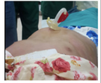
Gastrostomy tube in 8th day
- Connect the guide wire with the sharp head of the gastrostomy tube, carefully disinfect this connection before pulling the tube through the mouth (this is very important).
- Use the guide wire outside of the abdomen to gently pull the gastrostomy tube from mouth into stomach. When the sharp head of the tube touches the abdominal wall, using a knife lightly makes one small incision on the abdominal wall skin, it should be not too narrow and not too wide adequate with diameter of tube (20 or 24 fr).
- Continue gently pulling the tube from stomach until the bumper approaches the abdominal wall by using guide wire outside [1,3].
- The part of the tube outside the abdominal wall is fixed by external pumber to avoid deep movement of the tube into the stomach, while maintaining pressure between peritoneal and serosal membrane surface to create an adhesion [1, 3, 4].
- Endoscopy to check the tube position and to make sure no exceeding press on the abdominal wall [1, 3, 4].
- External bumpers will maintain standard pressure. Rotate the tube 90o in daily to reduce lesions and infiltration of mucosal stomach. Gastrostomy feeding can start 6 hours after procedure. Feeding tube can be shorter by cutting if it is too length [1].
After 12 weeks, fibrous tissue and mucosa automatically create adhesion between stomach and abdominal wall [1]. Feeding tubes can be replaced second times by other kinds of tube Bard (Bard MedicamVolketswil, Switzeland) with mushroom shape or MYC–KEY (Cosaum, Schlieren, Switzeland). These tools are more beautiful, more easy to hide under clothes and have a valve anti reflux to prevent skin lesions [1, 3].
After removing the gastrostomy tube or replaced tube, hole will close automatically after 48 hours. Sometimes, hole closes slowly, even two years later. In these cases, surgical closure is necessary.
REFERENCES
1. Gershman G (2012). “Therapeutic upper GI endoscopy”, Practical pediatric gastrointestinal endoscopy, (2), 82-103.
2. Victor LF (2008), “Gastrointestinal Endoscopy”, Pediatric gastrointestinal desase, 2(1), 1259-1348.
3. Mougenot J.F (2000), “Endoscopie digestive”, Gastroentérologiepédiatrique, (2), 664-685.
4. Phan ThịHiền (2017), “Mở thong dạ dày bằng nội soi”, Các qui trình kĩ thuật nhi khoa thường gặp, Nhà xuất bản Y học, 2, 364-365
Related posts
-

Self-design suction tool
20-05-2021 -

Removing phytobenzoar in Pig's stomach
20-05-2021 -

Remove twisting of the pig colon
04-05-2021 -

Pig stomach endoscopy
04-05-2021
-

Management of Ingested Foreign Bodies in Children: A Clinical Report of the NASPGHAN Endoscopy Committee
28-04-2021 -

Management of Familial Adenomatous Polyposis in Children and Adolescents: Position Paper From the ESPGHAN Polyposis Working Group
28-04-2021 -

Pediatric Colonoscopic Polypectomy Technique
28-04-2021 -

Gastrostomy Placement in Children: Percutaneous Endoscopic Gastrostomy or Laparoscopic Gastrostomy?
28-04-2021