Benign esophageal tumors
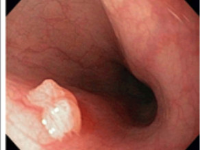
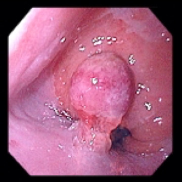
Squamous papilloma: verrucous papillated shape
.jpg)
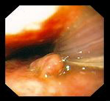
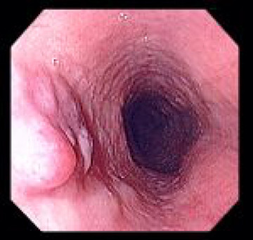
Leiomyoma: polypoid lesions
Fibrovascular Polyp
Malignant esophageal tumors
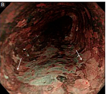
Squamous Cell Carcinoma: With narrow-band imaging, “metallic silver sign” , revealed mixture of stained and unstained area
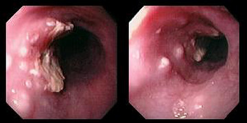
Multifocal Squamous Cell Carcinoma
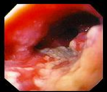
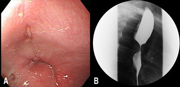
Adenocarcinoma: ulcerated, constricting tumor in several centimeters above the esophagogastric junction, arising in a segment of Barrett's esophagus
Initial management
•Biopsy: Histology examination
•Other test based on endoscopy images: esophagogram chest Xray; CT, endoscopic Ultrasonograph Bronchoscopy, PET scan
•Management:
–Endoscopic mucosal resection
–Radiotherapy
–Chemotherapy
–Surgery to remove part of or whole tumors
Related posts
- Normal esophagus - 03-05-2021
- Cytomegalovirus esophagitis - 04-05-2021
- Zenker's diverticulum (zd) - 29-04-2021
- Esophageal webs - 03-05-2021
- Gastric inlet patches in esophagus (heteropic gastric mucosa of the proximal esophagus) - 03-05-2021
- Esophageal glycoenic acanthosis - 03-05-2021
- Esophageal varices and sarin's classification for gastric varices - 03-05-2021
- Mallory - weiss tear - 03-05-2021
- Typical findings of primary esophageal achalasia - 03-05-2021
- Esophageal stenosis - 03-05-2021
-

Self-design suction tool
20-05-2021 -

Removing phytobenzoar in Pig's stomach
20-05-2021 -

Remove twisting of the pig colon
04-05-2021 -

Pig stomach endoscopy
04-05-2021
-

Management of Ingested Foreign Bodies in Children: A Clinical Report of the NASPGHAN Endoscopy Committee
28-04-2021 -

Management of Familial Adenomatous Polyposis in Children and Adolescents: Position Paper From the ESPGHAN Polyposis Working Group
28-04-2021 -

Pediatric Colonoscopic Polypectomy Technique
28-04-2021 -

Gastrostomy Placement in Children: Percutaneous Endoscopic Gastrostomy or Laparoscopic Gastrostomy?
28-04-2021