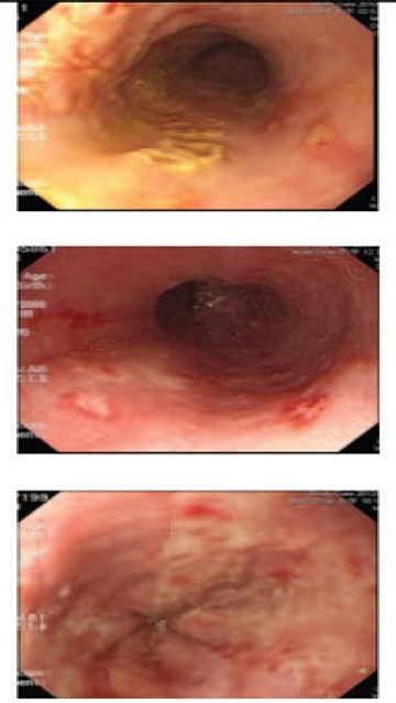
Classification of herpes esophagitis of itoh
Type 1: The middle and lower thirds of the esophagus: Small, punched-out lesions with raised margins; a slightly yellowish color and fibrin exudation at the center of lesions
Type 2: The middle and lower thirds of the esophagus: Small, punched-out lesions without raised margins; no color and no exudation
Type 3: Multiple ulcers become confluent, like a map at the extended esophageal lesions

Initial treatment
•Biopsy: Histological identification of HSV for ∆+
•Blood tests: Serological HSV anti IgM, anti-HSV IgG
•Others test: Immunodeficiency, renal failure, malignancy, transplanted organs, HIV, steroid or immunosuppressive therapy
•Therapy: Antivirus (Acyclovir, Famciclovir, Valacyclovir)
Related posts
- Normal esophagus - 03-05-2021
- Cytomegalovirus esophagitis - 04-05-2021
- Zenker's diverticulum (zd) - 29-04-2021
- Esophageal webs - 03-05-2021
- Gastric inlet patches in esophagus (heteropic gastric mucosa of the proximal esophagus) - 03-05-2021
- Esophageal glycoenic acanthosis - 03-05-2021
- Benign and malignant esophageal tumors - 03-05-2021
- Esophageal varices and sarin's classification for gastric varices - 03-05-2021
- Mallory - weiss tear - 03-05-2021
- Typical findings of primary esophageal achalasia - 03-05-2021
EDUCATION
-

Self-design suction tool
20-05-2021 -

Removing phytobenzoar in Pig's stomach
20-05-2021 -

Remove twisting of the pig colon
04-05-2021 -

Pig stomach endoscopy
04-05-2021
Recommendation
-

Management of Ingested Foreign Bodies in Children: A Clinical Report of the NASPGHAN Endoscopy Committee
28-04-2021 -

Management of Familial Adenomatous Polyposis in Children and Adolescents: Position Paper From the ESPGHAN Polyposis Working Group
28-04-2021 -

Pediatric Colonoscopic Polypectomy Technique
28-04-2021 -

Gastrostomy Placement in Children: Percutaneous Endoscopic Gastrostomy or Laparoscopic Gastrostomy?
28-04-2021
Videos
Contact


