Esophagus
27-04-2021
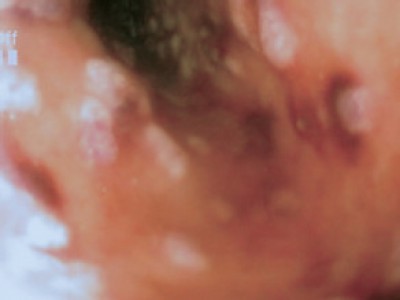
The images of pathological esophagus (translation)
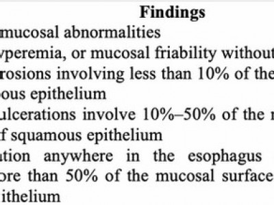
The images of abnormal esophagus are detected by endoscopy mostly related to specific pathology. In which, a common disease is gastroesophageal reflux (GER), especially in children with breast feeding. This disease can lead to ulcerative esophagus, even esophageal...
27-04-2021
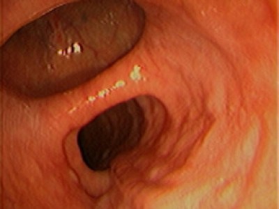
Esophageal and cardia dilatation (translation)
Phan Thi Hien, "Esophagus-stomach-duodenum endoscopy in children", Medical Publishing House, Hanoi, Jan 2019 (page 125-137). Extract from specialty book: “NỘI SOI THỰC QUẢN – DẠ DÀY – TÁ TRÀNG TRẺ EM”, Nhà xuất...
23-01-2021
Management of bleeding without esophageal varices (translation)
Phan Thi Hien, "Esophagus-stomach-duodenum endoscopy in children";, Medical Publishing House, Hanoi, Jan 2019 (page 151-163). Extract from specialty book: “NỘI SOI THỰC QUẢN – DẠ DÀY – TÁ TRÀNG TRẺ EM”, Nhà xuất...
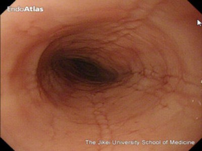
03-05-2021
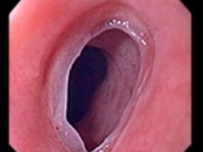
Normal esophagus
Z-line: The junction between the pale esophageal and richer colored gastric mucosa is slightly irregular. It is located at the level or within 2cm above the hiatal notch.
04-05-2021
Cytomegalovirus esophagitis
Lesion: Esophageal ulceration and vasculitis caused by CMV infection at multiple sites
Biopsy: ≥ 10 specimens from the base of the ulcer. ∆+: Histology identification of CMV by immunohistochemical and direct fluorescence...
29-04-2021
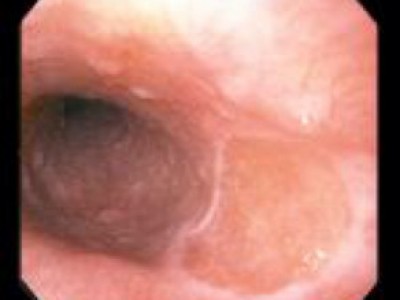
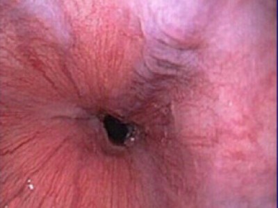
Zenker's diverticulum (zd)
Lesion: Sac-like outpouching of the mucosa and submucosa layers located dorsally at the pharyngoesophageal
03-05-2021
Esophageal webs
Lesion: Smooth, circumferential ring of squamous mucosa, which can be located anywhere along the esophagus
03-05-2021
Gastric inlet patches in esophagus (heteropic gastric mucosa of the proximal esophagus)
Lesion: salmon-colored patch of mucosa in the proximal esophagus, just below the upper esophageal sphincter. This is an island of heterotopic gastric mucosa, appears distinct from the surrounding squamous mucosa, which has a silvery color*, with 0,8cm...
03-05-2021
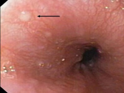
Esophageal glycoenic acanthosis
Lesion: in the distal esophagus, as characterized by multiple small, raised pale nodules, generally benign mucosal lesions
03-05-2021
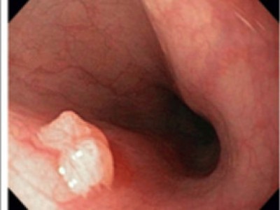
Benign and malignant esophageal tumors
Squamous papilloma: verrucous papillated shape
Leiomyoma: polypoid lesions
03-05-2021
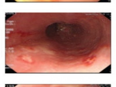
Esophageal varices and sarin's classification for gastric varices
Grade 1: Varices disappear with air insufflation
Grade 2: Non-confluent varices remain identical with air insufflation
03-05-2021
Mallory - weiss tear
Lesion: Bleeding from a laceration in the mucosa at the junction of the stomach and esophagus
03-05-2021
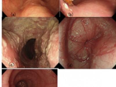
Typical findings of primary esophageal achalasia
(a) Dilation of the esophagus. Dilated esophagus drooped to both sides of the spine.
(b) Food remnant in the esophagus
(c) Whitish coating of the mucosa caused by adhesion of the remained food inside of the...
03-05-2021
Esophageal stenosis
Lesion: Esophageal stenosis (1mm of diameter) at 4cm from upper sphincter and 13cm from front teeth, straight shaft, no oedema, no diverticula, no fistula in the boy 2-year-old operated esophagael astresia

03-05-2021
Zagar classification for corrosive ingestion
(A) Grade 1 indicates only slight swelling and redness of the mucosa.
(B) Grade 2A indicates the presence of superficial ulcers, bleeding, and exudates.
(C) Grade 2B indicates local or encircling deep...
03-05-2021
Classification of herpes esophagitis of itoh
Type 1: The middle and lower thirds of the esophagus: Small, punched-out lesions with raised margins; a slightly yellowish color and fibrin exudation at the center of lesions
Type 2: The middle and lower thirds of the esophagus:...
03-05-2021
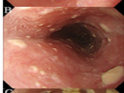
The severity of esophageal candidiasis (ce) according to kodsi's classification
Grade I: a few raised white plaques up to 2 mm in size without edema or ulceration
Grade II: multiple raised white plaques greater than 2 mm in size without ulceration
Grade III: confluent, linear,...
03-05-2021
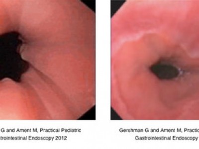
Eosinophilic esophagitis
Lesion: 1/2 under the esophagus, longitudinal furrowing/shearing and the “crêpe paper”
Alberto Ravelli, Practical pediatric gastrointestinal endoscopyractical, 2012
04-05-2021
Hiatus hernia according to the modified makuuchi classification (hh)
Hiatus hernia according to the modified Makuuchi classification (HH)
03-05-2021
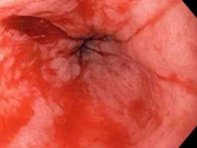
Esophageal inflammatory polyp-fold complex
Lesion: Endoscopic view of an esophageal inflammatory polyp-fold complex in an 8-year-old with recalcitrant gastroesophageal reflux
Trang: 1 | 2
EDUCATION
-

Self-design suction tool
20-05-2021 -

Removing phytobenzoar in Pig's stomach
20-05-2021 -

Remove twisting of the pig colon
04-05-2021 -

Pig stomach endoscopy
04-05-2021
Recommendation
-

Management of Ingested Foreign Bodies in Children: A Clinical Report of the NASPGHAN Endoscopy Committee
28-04-2021 -

Management of Familial Adenomatous Polyposis in Children and Adolescents: Position Paper From the ESPGHAN Polyposis Working Group
28-04-2021 -

Pediatric Colonoscopic Polypectomy Technique
28-04-2021 -

Gastrostomy Placement in Children: Percutaneous Endoscopic Gastrostomy or Laparoscopic Gastrostomy?
28-04-2021

Videos


Contact